Wild-Type Amyloidosis
The Role of Imaging in Amyloidosis Diagnosis & Management
A heart specialist and leader of OHSU’s Cardiac Amyloidosis and Hypertrophic Cardiomyopathy programs, Dr. Masri discussed how imaging plays a key role in identifying amyloidosis and monitoring treatment.
Read More
AL Amyloidosis
ATTR Amyloidosis Clinical Trials (January 2026)
Kristen Hsu, ARC’s Executive Director of Research, shared the latest updates on ATTR (transthyretin amyloidosis) clinical trials and what these developments mean for patients and families. There was also an opportunity for questions and answers.
Read More
Webinars
Coffee Chat with ARC 2025
This webinar “Coffee Chat” style webinar is your chance to learn about the different programs and projects ARC has been working on in 2025! Hear directly from ARC staff how we are supporting patients and helping shape the future of amyloidosis.
Read More
AL Amyloidosis
Tips for Surviving & Thriving as a Care Partner
Clinical Psychologist Rosalind Kalb, PhD shared helpful advice, self-care tips, and real-life strategies to make daily life a little easier for both Care Partners and loved ones. Linnie, an amyloidosis Care Partner, also shared experiences and perspectives from her own life.
Read More
AL Amyloidosis
Neuropathy and Amyloidosis
Dr. Sami Khella, Professor of Neurology and co-founder of the Amyloidosis Program at the University of Pennsylvania discussed the latest in diagnosis, treatment, and management of neuropathy and amyloidosis.
Read More
Webinars
ATTR Clinical Trial Updates
ARC Executive Director of Research Kristen Hsu presented an overview of the different approaches to treating ATTR amyloidosis, as well as the most current updates for the clinical trial landscape, with a focus on ATTR trials.
Read More
Hereditary ATTR Amyloidosis
ATTR Cardiac Treatments
Dr. Jan Griffin, amyloidosis cardiologist at the Medical University of South Carolina, discussed how ATTR affects the heart and explored advances in treatments for ATTR-CM.
Read More
Webinars
ARC Research Roundtable Report
This white paper summarizes advancements in the field, key challenges, and consensus priorities for the amyloidosis research community that were identified and discussed by attendees of the 2024 ARC Research Roundtable Meeting. The information presented in this paper represents the opinions and outlooks of the global amyloidosis experts.
Read More
AL Amyloidosis
Understanding Living with Amyloidosis: Insights from the ARC Annual Community Survey
Sabrina Rebello, ARC's Senior Research Manager, presented an in-depth overview of key insights from our Annual Community Survey. This webinar focused on understanding the experiences of patients and caregivers throughout their diagnostic journey and ongoing care.
Read More
AL Amyloidosis
Reliability and validity of the Transthyretin Amyloidosis – Quality of Life (ATTR-QOL) Questionnaire impact scales
Lovley, A., Hsu, K., LaGasse, K. et al. Reliability and validity of the Transthyretin Amyloidosis – Quality of Life (ATTR-QOL) Questionnaire impact scales. J Patient Rep Outcomes 9, 44 (2025). https://doi.org/10.1186/s41687-025-00880-7
Visit Website
Hereditary ATTR Amyloidosis
Emotional Aspects of Amyloidosis
Dr. Meghan Beier, psychologist at the Rowan Center for Behavioral Medicine, explored the emotional aspects of amyloidosis. She discussed mental health issues in amyloidosis and present strategies for coping.
Read More
AL Amyloidosis
Development of Imaging Endpoints for Clinical Trials in AL and ATTR Amyloidosis: Proceedings of the Amyloidosis Forum
Dorbala, S, Adigun, R, Alexander, K. et al. Development of Imaging Endpoints for Clinical Trials in AL and ATTR Amyloidosis: Proceedings of the Amyloidosis Forum. J Am Coll Cardiol Img. null2025, 0 (0) .
https://doi.org/10.1016/j.jcmg.2024.11.003
Visit Website
AL Amyloidosis
Amyloidosis 101
Dr. Sarah Cuddy, cardiologist at Brigham and Women's Hospital, explored the basics of amyloidosis. She presented an introduction to amyloidosis, the different types, and the available treatments. The webinar concluded with a Q&A session.
Read More
AL Amyloidosis
Factors associated with financial toxicity in patients with transthyretin amyloidosis: results from Amyloidosis Research Consortium’s treatment affordability patient and caregiver survey
Rebello, S., Hsu, K., Nativi-Nicolau, J., Karam, C., Grogan, M., Lousada, I., & Maurer, M. S. (2025). Factors associated with financial toxicity in patients with transthyretin amyloidosis: results from Amyloidosis Research Consortium’s treatment affordability patient and caregiver survey. Amyloid, 1–10. https://doi.org/10.1080/13506129.2025.2462541
Visit Website
Hereditary ATTR Amyloidosis
Clinical Trial Updates for the ATTR Community
ARC Executive Director of Research Kristen Hsu presented the most current updates for the clinical trial landscape, with a focus on ATTR amyloidosis. Kristen will discussed the direction of future research. The webinar concluded with a Q&A session.
Read More
Hereditary ATTR Amyloidosis
Coffee Chat with ARC
To wrap up 2024, this month’s ARC Talks Webinar consisted of a conversation with ARC staff. We shared highlights from 2024, started to look ahead to 2025, and answered your questions about the work we do for the amyloidosis community.
Read More
AL Amyloidosis
Caring for the Caregiver
In honor of National Family Caregivers Month, ARC hosted a discussion between Debra Ruehlman, amyloidosis family caregiver, and Ann Payne, MSW, ARC Clinical Care and Education Manager. This webinar explored topics such as self care, self advocacy, and community resources.
Read More
AL Amyloidosis
Medicare and the Evolving Financial Landscape
Covering information on Medicare basics, choosing a Part D plan, and coverage gaps, ARC was joined by Sylvia Gary from the Centers for Medicare and Medicaid Services (CMS).
Read More
AL Amyloidosis
GI Disturbances and Symptom Management
Dr. Sara Horst discussed how amyloidosis can impact the gastrointestinal tract, GI symptoms, and symptom management.
Read More
AL Amyloidosis
Clinical Trial Updates for the ATTR Community
ARC's own Kristen Hsu presented the most current updates for the clinical trial landscape, with a focus on ATTR amyloidosis.
Read More
Hereditary ATTR Amyloidosis
Physical Therapy for Amyloidosis Management
Physical therapists Kelsi Schiltz, and Katie Johnson provided recommendations for managing neuropathy symptoms.
Read More
AL Amyloidosis
Visualizing Amyloidosis: The Role of Imaging
Dr. Sharmila Dorbala explained the role that medical imaging techniques play in both diagnosing and managing amyloidosis.
Read More
AL Amyloidosis
Devices for Heart Improvement
Amyloidosis cardiologist, Dr. Mazen Hanna discusses the function of implantable, electronic devices in cardiac management of amyloidosis patients.
Read More
AL Amyloidosis
An ARC Fireside Chat
The dedicated team behind the transformative initiatives of ARC discuss the impactful work of the Leadership, Research, Development, Communications, and Community Alliance departments.
Read More
AL Amyloidosis
Navigating Care Choices at the End of Life
Jill Waldron, NP and Rebecca Kirch provide guidance on navigating care choice options for amyloidosis patients and caregivers during health decline.
Read More
AL Amyloidosis
Knowing Your Rights: Understanding Health insurance Coverage
Alicia Lawrence, Information and Resource Services Manager at the National Organization for Rare Disorders (NORD), discussed health insurance policies within private and government issued insurance.
Read More
AL Amyloidosis
Kidney Disease Presentation in Amyloidosis
Dr. Nelson Leung focused on renal involvement in AL, AA, ALECT2 and ATTR amyloidosis, while discussing treatment and response assessments for each type.
Read More
Webinars
Cardiac and Neurological Approaches in ATTR Management
Dr. Nativi-Nicolau and Dr. Elizabeth Mauricio discussed the decision pathway in management of ATTR amyloidosis from the cardiac and neurological standpoint.
Read More
Webinars

Wild-type ATTR: Ralph’s story
Ralph, a patient with Wild-Type ATTR amyloidosis, describes his journey to diagnosis. A commercial and technical diver, Ralph’s bilateral carpal tunnel was often attributed to his career. After multiple doctors misdiagnosed Ralph, one physician sent a tissue sample out for a biopsy, which led to Ralph’s amyloidosis diagnosis. From there, a journey of self-education, empowerment, and treatment have helped him take on amyloidosis and keep living, despite the challenges of this difficult disease.
Read More
Patient Stories
Overview of ARC’s Research Programs and Results of the Patient Community Survey
Kristen Hsu, Executive Director of Research, and Sabrina Rebello, Research Manager, provide an overview of ARC’s research programs, share findings from our 2022 community survey results, and highlight the future direction of ARC’s research.
Read More
Hereditary ATTR Amyloidosis
Current ATTR landscape: Treatments and Trials
Amyloidosis expert Dr. John L. Berk presents the most current treatment options for ATTR amyloidosis. Dr. Berk also discusses clinical trials and future research. The webinar concludes with a Q and A session.
Read More
Hereditary ATTR Amyloidosis
Amyloidosis and Nutrition: Eating for Your Health
Briana Pineau, MS, RD, LDN, a registered dietician at Boston Medical Center, discusses the role nutrition can play in the management of amyloidosis symptoms. In addition to covering gastrointestinal involvement associated with various types of amyloidosis, Briana covers the broader health benefits of a balanced diet.
Read More
AL Amyloidosis
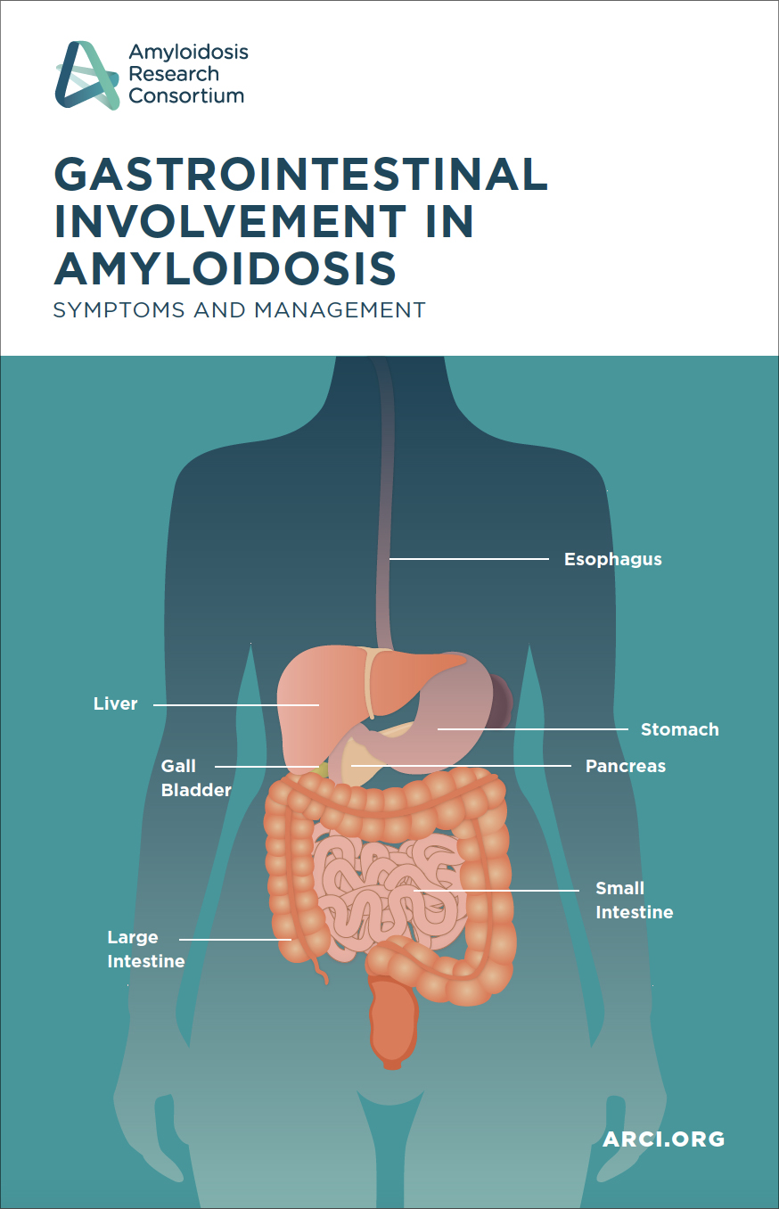
Gastrointestinal Involvement in Amyloidosis
Many types of amyloidosis can affect patients’ gastrointestinal (GI) tract, causing painful, annoying, or even debilitating symptoms. It is common for patients with amyloidosis to experience GI involvement and in fact, some patients may have GI-related symptoms as their most predominant sign or chief complaint.
Read More
AL Amyloidosis
The Heart of the Matter: Cardiac Amyloidosis
Dr. Kevin Alexander, a cardiac amyloidosis specialist from Stanford University, discusses the cardiac signs and symptoms to recognize, treatment and management options for each type of Amyloidosis and the future direction of research in the field.
Read More
AL Amyloidosis
Empowering Patients: Navigating Your Care
Isabelle Lousada shares her experiences with amyloidosis and the lessons learnt on how to become an empowered and informed patient. Lisa Mendelson, nurse practitioner from Boston University’s Amyloidosis Program provides a valuable medical perspective about how to build a successful relationship with your care team. Original presentation date June 23, 2022.
Read More
AL Amyloidosis
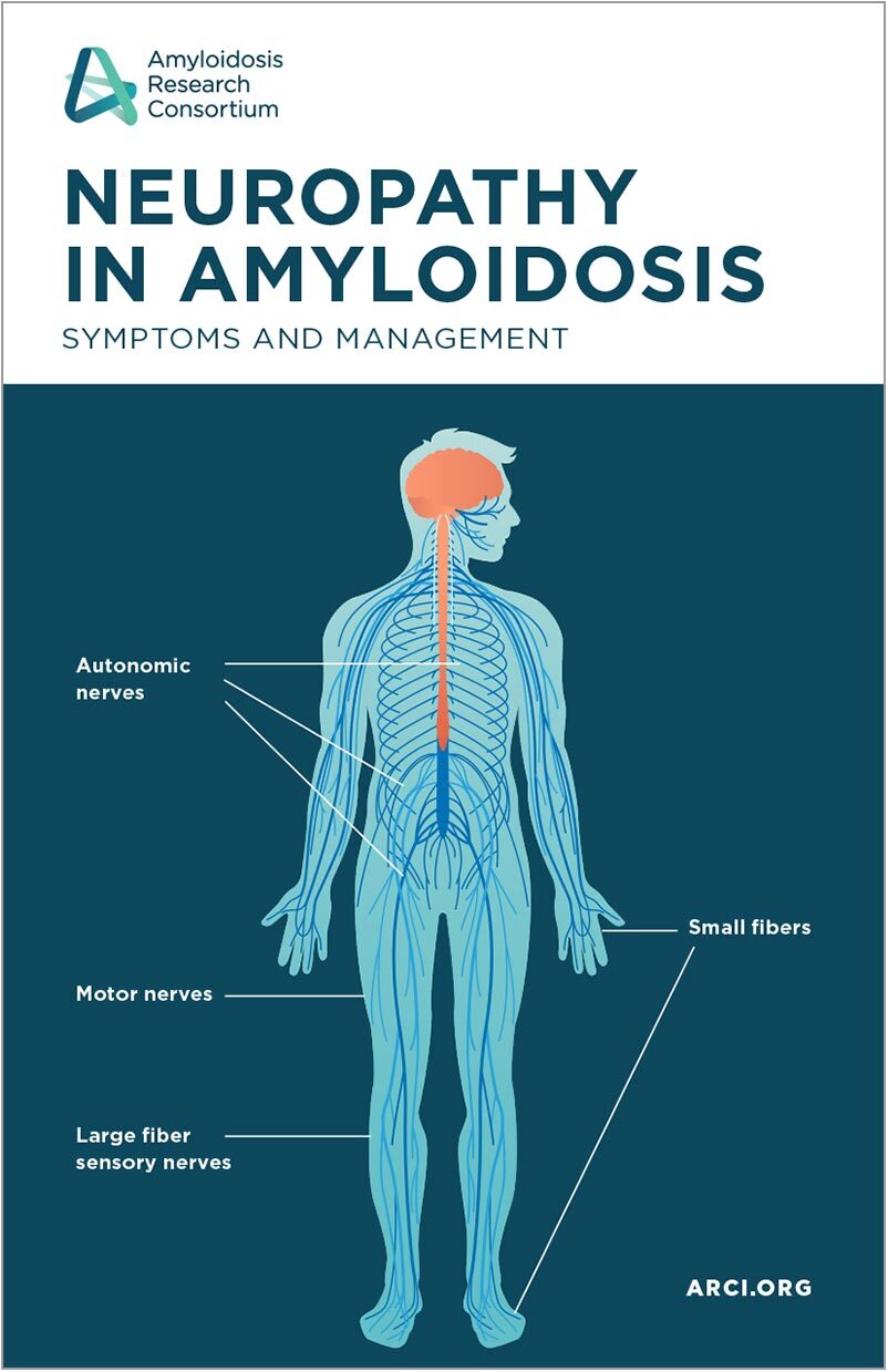
Neuropathy in Amyloidosis
Many types of amyloidosis can affect patients’ nerves, causing painful, annoying, or even debilitating symptoms. It is common for patients with amyloidosis to experience neuropathy and in fact, some patients may have nerve-related symptoms as their most predominant sign or chief complaint.
Read More
AL Amyloidosis
Mental Health Matters: Caring for your Wellbeing
Rare disease mental health expert Kym Winter will equip you with the resources and tools you need to take care of your mental health.
In this interactive session, you will be introduced to the Stress Bucket Approach, which is a simple way of thinking about and looking after your own - and others’ - emotional wellbeing in order to live well with the impacts of a rare disease such as amyloidosis. Original presentation date February 23, 2022
Read More
AL Amyloidosis
Coffee with ARC: Learn about ARC’s work
In this ARC Talks special presentation, Coffee with ARC, some members of the ARC team will provide an overview of ARC's history and areas of focus, as well as our plans for 2022 and beyond. Original presentation date December 9, 2021
Read More
AL Amyloidosis
Living Well with Amyloidosis
In amyloidosis, common symptoms such as gastrointestinal manifestations and neuropathy are often the most troublesome for patients. In our ARC Talks Webinar for patients and caregivers, amyloidosis experts from across multiple specialties explain approaches for symptom management. Our experts provide you with the knowledge you need to live well with amyloidosis. Original presentation date October 26, 2021
Read More
AL Amyloidosis
Taming Wild-Type
This ARC Talks Webinar covers everything that patients and their families should know about wild-type transthyretin amyloidosis. Dr. Martha Grogan, Founder and Director of the Cardiac Amyloid Clinic at the Mayo Clinic, covers disease progression, prognosis, treatment options, and more. Dr Grogan then answers questions from the audience from our live presentation on June 10, 2021.
Read More
Wild-Type Amyloidosis
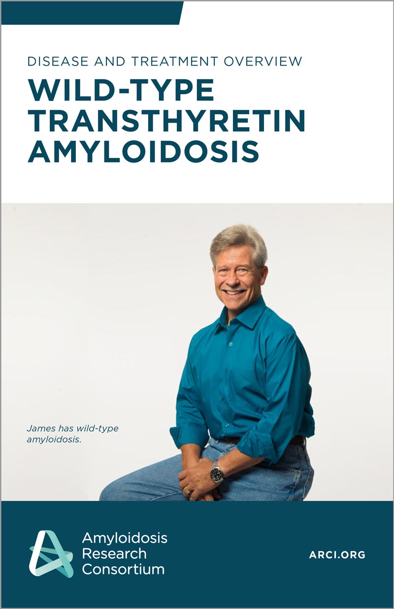
Disease and Treatment Overview: Wild-Type Transthyretin Amyloidosis
Wild-type transthyretin amyloidosis (ATTRwt) is an age-related disease caused by transthyretin (TTR) proteins that become unstable, misfold, and build up in organs, impairing their function. It is a slowly progressive condition that affects older people, most often Caucasian men over 65 years of age. Heart disease is the hallmark of ATTRwt, but it is commonly preceded by other conditions, such as carpal tunnel syndrome or spinal stenosis.
Read More
Library
Introducción a la Enfermedad y Tratamiento: Amiloidosis Por Transtirrentia Natural
La amiloidosis por transtiretina de tipo salvaje (ATTRwt) es una enfermedad relacionada con la edad causada por proteínas transtiretina (TTR) que se vuelven inestables, se pliegan mal y se acumulan en los órganos, lo que altera su función. Es una afección de progresión lenta que afecta a personas mayores, con mayor frecuencia a hombres caucásicos mayores de 65 años. La enfermedad cardíaca es el sello distintivo de ATTRwt, pero comúnmente va precedida por otras afecciones, como el síndrome del túnel carpiano o la estenosis espinal.
Read More
Library
Amyloid Neuropathy Burden and Mangement
In this ARC Talks webinar, Dr. Kelsey Barrell from the University of Utah explains what causes neuropathy and other neurological symptoms in amyloidosis patients and offers suggestions to help manage these symptoms.
Read More
AL Amyloidosis
Caregivers: A Guide to Self-Care
Nancy Verel, a nurse at the Cleveland Clinic, shares her story about her family's journey through her husband's AL amyloidosis diagnosis and treatment. Robert David from BMC Cancer Support Programs provides coping and support strategies for caregivers.
Read More
AL Amyloidosis
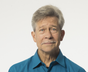
Wild-type ATTR: James’ story
James' only concerning symptom leading up to his diagnosis was bilateral edema. But, after a stroke, James spent a month in the hospital and received a surprising diagnosis of Wild-Type Transthyretin Amyloidosis. James shares his story and optimism just months after his diagnosis.
Read More
Patient Stories
Physical and Occupational Therapy – Managing Your Amyloidosis
In this patient webinar, Mayo Clinic's occupational therapist Sarah Dahlhauser, OTD, OTR/L, and physical therapist Sarah Boyd, PT, DPT, discuss exercise principles for maintaining mobility and function, and home modifications for improved safety for amyloidosis patients.
Read More
AL Amyloidosis
Webinar: What is Expanded Access?
This webinar covers access to innovative new therapies through expanded access programs with guest speakers Jennifer Miller, PhD, Assistant Professor at Yale University School of Medicine and Alison Bateman-House, PhD, MPH, Assistant Professor at NYU School of Medicine.
Visit Website
AL Amyloidosis
No Resources found for the chosen category